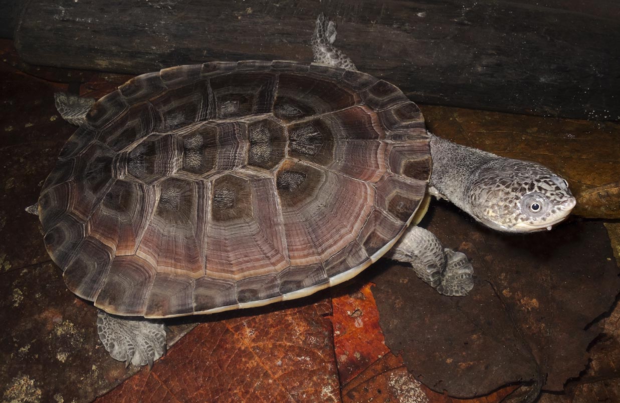
Melanopsin mRNA in the Iris of Red-Eared Slider Turtles (Trachemys scripta elegans)
The iris in Red-Eared Slider Turtles (Trachemys scripta elegans) is intrinsically photosensitive, slowly constricting the pupil when exposed to light. We hypothesized that one of the photopigments involved in the photomechanical response (PMR) is melanopsin (Opn4). The purpose of this project was to determine whether mRNA of Opn4 is present in their iris. After eyes were dissected from animals, we extracted iris tissues to carry out fluorescent in situ hybridization. Comparison of fluorescence from images revealed significant labeling in the iris for two melanopsin isoforms (Opn4x and Opn4m). We then carried out quantitative real-time PCR to identify relative expression levels of mRNA. Expressions by both isoforms in the iris were significant. In contrast to the retina, which had higher levels of Opn4m than Opn4x, the iris had higher levels of Opn4x than Opn4m. We also carried out Carr–Price reactions on tissue extracts, which were stabilized by hydroxylamine, and detected retinal oxime in the iris. Possession of retinal oxime indicates prior presence of retinal, a vitamin A-type chromophore, necessary for a functional protein. Evidence of a chromophore along with both Opn4 mRNA isoforms in the iris of this species supports presence of the photopigment and a possible role in the turtles' slow PMR.Abstract

Alignment of Opn4 transcripts in turtles. (A) Partial cds (coding DNA sequences) for Opn4x and Opn4m of Red-Eared Slider Turtles with predicted cds for Western Painted Turtles. The predicted Opn4x mRNA is 1,795 bp composed of 10 exons (cds: 32–1,708 bp, i.e., length = 1,677 bp); the Opn4m mRNA is 4,551 bp, 11 exons (cds = 1–1,623 bp). (B) Transcripts were computed from whole shotgun genomic sequences submitted to the National Center for Biotechnology (NCBI) by the Painted Turtle Genome Sequencing Consortium—The Genome Institute at Washington University (Shaffer et al., 2013; Badenhorst et al., 2015). The Opn4x gene is located on an unplaced scaffold 1,788,267 to 1,857,788 bp; length = 69,522 bp (Reference Sequence: NW_007359856); the loci for Opn4m also on unplaced scaffold is from 1,582,394 to 1,610,018 bp; length = 27,625 bp (Reference Sequence: NW_007281381). Noncoding DNA sequences are in gray; location is at end of Opn4m and given smaller portions barely noticeable at front and back for Opn4x. (C) The Opn4x sequences had 99% similar identity (467/473, 1 gap); the Opn4m sequences 99% similar (988/995). Alignments of Opn4x to Opn4m also were similar, which further confirmed NCBI predictions. For example, the Opn4x of Red-Eared Slider Turtles showed 67% identity (241/362, 17 gaps) to the Opn4m of Western Painted Turtles; the Opn4x of Western Painted Turtles also 67% identity (506/755, 18 gaps) to their Opn4m.

Translations of 472 and 1,004 bp partial transcipts for Opn4x and Opn4m, respectively, of Red-Eared Slider Turtles aligned with predicted versions for Western Painted Turtles. The 157 amino acid translation of the Opn4x aligned at 100% identity (144/144, 91% query cover) with the 558 amino acid sequence predicted for the Opn4x form in Western Painted Turtles. The partial sequence of the Opn4m form aligned with 99% identity (331/334) to the 540 amino acid sequence predicted for Western Painted Turtles. Predictions for the locations of seven transmembrane domains were done using TMPred (http://www.ch.embnet.org/ software/TMPRED_form.html) (Artimo et al., 2012) and are shown with horizontal bars: green for Opn4x and red for Opn4m. Amino acids surrounded by black boxes are common features for the family of Opn4, that is, two cysteine (C) residues for disulfide bond formation, the aspartate/arginine/tyrosine (D/R/Y) tripeptide for binding transducin, tyrosine (Y) and glutamate (E) for counterion sites, and lysine (K) for the Schiff's base (Bellingham et al., 2006). Residues in gray highlight are those conserved among the sequences. Of the residues conserved beyond the seventh transmembrane span and located in the carboxy terminal tail, a large proportion are either serine or threonine, 36% (15/42) (yellow highlight). These amino acids are potential sites of phosphorylation and, therefore, could be important for deactivation of the protein via kinases and/or interaction with arrestin (Panda et al., 2005; Tobin, 2008; Blasic et al., 2014) (see Discussion).

Confocal and SEM images compared at low magnification to provide context for the labeleling by probes. Images (0.805 pixel/μm) of the posterior surface of an iris labeled with (A) Opn4x and (B) Opn4m antisense probes. Lower right quadrants of A and B are images from an iris treated with sense probes (control). Images were captured by a Nikon A1 confocal microscope (10×/0.45 numerical aperture (NA) Plan Apochromat objective). FAM Opn4x and ATTO550N Opn4m probes were excited by 488 and 561 nm lasers and viewed with filters 525 ± 25 and 595 ± 25 nm, respectively. Tiles were stitched together to generate montages using Nikon's NIS-Elements software. (C) Magnified (two times) image of Opn4m labeling overlaid onto Opn4x to show colocalization of hybridization. The rectangle with yellow border frames a region of the dilator pupillae, that is, radial structure (within yellow arrow heads); a rectangle with purple border frames an area of the sphincter pupillae, that is, circular organization (within purple arrow heads). The asterisk identifies where the tissue pattern begins to change from being radial to circular. (D) Scanning electron micrograph (0.192 pixels/μm) of the posterior surface of another iris of similar size and shape (scale is same as in C). Rectangles with white border identify regions of the dilator pupillae and sphinctor pupillae, which correspond to similar regions shown in C. The radial pattern is clearly visible for the dilator pupillae (yellow arrow heads) along with a location (asterisk) marking the transition to a pattern that becomes less so toward the pupil.

Regions of the confocal images shown in Figure 3C to highlight features of the iris. (A) The dilator pupillae is the organization of structures radiating outward (yellow borders). Transition from a radial to circular pattern is identified with an asterisk. (B) The sphincter pupillae is defined by cells arranged in concentric circular patterns around the circumference of the pupil (purple borders). Left column is colocalization; middle column is labeling by Opn4x; and right column by Opn4m.

Antisense stained images showed greater fluorescence than sense and blanks. Values of corrected total cell fluorescence (CTCF) were calculated from dorsal, ventral, temporal, and nasal regions (gray cartoon of iris) and normalized to the maximum found among the regions in treated tissues. Regions were selected so that they included portions of both the dilator pupillae and the sphincter pupillae. (A) Comparison of means ± SE from three sets of irises (antisense, sense, and blank) measured for Opn4x (green) and Opn4m (red) labeling using a Nikon E800 C1 confocal laser scanning microscope (20×/0.75 NA Plan Apochromat objective). FAM Opn4x probes were excited by the 488 nm laser and viewed with a 515 ± 15 nm filter; ATTO550N Opn4m probes were excited by the 543 nm laser and viewed with a 595 ± 25 nm filter. Asterisk denotes significant difference of antisense from both sense and blank. (B) Comparison from another three sets of irises (antisense and sense) measured using an Olympus FV10i confocal laser scanning microscope (10×/0.40 NA UPLSA Apo objective). Opn4x and Opn4m probes were excited by 473 and 559 nm lasers and viewed with filters 519 ± 30 and 603 ± 30 nm, respectively. As in (A), antisense was significantly different than sense (asterisk). Although SE bars for antisense Opn4x and Opn4m are nonoverlapping, the difference was not significant.

Confirmation of hybridization in epifluorescent images (4.56 pixel/μm) from a set of tissues at high magnification (40×/1.30 NA Oil Plan Fluor objective) using the Nikon E800 C1: irises treated with antisense probes (A) and sense probes (B); also included is a blank (C). DAPI stain was excited by 393 ± 8 nm and viewed with emission filter 458 ± 8 nm. Opn4x and Opn4m probes were excited by 480 ± 15 and 535 ± 25 nm, and viewed with 535 ± 20 and 600 ± 20 nm, respectively. Images were captured by a QIClick™ color CCD camera. Location of images were at a central portion of the dorsal region of irises, one of the areas used for determining intensity of fluorescence by scans done with confocal microscopes (small rectangle with white border in gray cartoon of an iris). In irises treated with antisense probes (A), nuclei are clear and individual Opn4x positive cells can be seen at locations on either side of the spoke of the dilator pupillae. Brighter yellow cells located on the spoke and in the sphinctor in A (colocalization) compared to those in B and C also indicate positive labeling for both Opn4x and Opn4m. Colocalization of images for green (Opn4x) and red (Opn4m) channels (right column) was done with Adobe Photoshop using the overlay blending mode.

Levels of Opn4x and Opn4m gene expression in the iris relative to the retina. (A) Threshold cycle (Ct) plotted vs. serial dilutions of iridal cDNA for each primer pair showed acceptable efficiencies and slopes: Efficiency = {[10(−1/slope)]−1} × 100%. For the Opn4x primer pair, efficiency = 106.3%, slope = −3.72, and r2 = 0.98; for the Opn4m pair, efficiency = 108.2%, slope = −3.14, and r2 = 0.98. (B) Melting curves of amplicons from the serial dilutions have isolated peaks, Opn4x (green traces) at 80°C and Opn4m (green traces) at 81°C. RFU = relative fluorescent units. (C) An example of melting curves of amplicons from one set of tissues: iris, retina, and liver. Peaks for Opn4x (green traces) and Opn4m (red traces) were at the expected temperatures. Peak for house-keeping, β-actin (black traces) was 84.5°C. Because each sample was done in triplicate, traces are N = 9 for each amplicon type. (D) Analysis of amplicons from iridal samples on a 2% agarose, 1× tris-acetate buffer gel. Sizes were as expected: 75 bp for Opn4x, 99 bp for Opn4m, and 96 bp for β-actin. (E) Bar plot shows mean levels of Opn4x and Opn4m expression in the iris and the retina normalized to liver. Vertical error bars in plot are ± 95% confidence limits (CL). Liver values for Opn4x and Opn4m were 1.0 ± 0.28 (± CL) and 1 ± 0.97, respectively (because of scale differential, they are not plotted).

Spectrophotometry revealed presence of retinal oxime in extracts of the iris and pineal first stabilized with hydroxyalmine, then reacted with Carr–Price reagent. (A) Peaks for standards in plots of absorbance in units of optical density (O.D.) vs. wavelength (λ) in nm. Amounts reacted were 3.4 nmole (retinyl palmitate, blue trace), 12 nmole (retinal, blue-green trace), and 24 nmole (retinal oxime, purple-brown trace). Colors of traces are those that we observed immediately after combining. Molar absorption coefficients (ɛ) in units of M−1·cm−1 confirmed reactions. Retinyl palmitate was 132.4 (λmax = 620.5 nm), −0.5% of accepted value (Baxter and Robeson, 1942). Retinal was 100.9 (λmax = 663 nm), +2% (Robeson et al., 1955). Retinal oxime, 69.43 (λmax = 595 nm), fell within the range, 44 to 113, identifiable for retinyl cation, an intermediate species in Carr–Price reactions with vitamin A-containing solutions (Blatz and Pippert, 1968; Blatz et al., 1971; Blatz and Estrada, 1972; Kiser et al., 2014). A lesser but noticeable hump also was apparent at 664 nm. After 3 min, these disappeared within an absorbance that rose toward a longer lasting peak of 480 nm (see Pitt et al., 1955). (B) Values are plotted for tissue extracts. Peak for iris was 593; peak for pineal 598 nm. The retina possessed a primary peak at 696 nm and secondary at 608 nm; the liver possessed humps also at these locations, but the order of the magnitude was vice versa. Colors of traces again are those observed after reactions: purple-brown (iris); yellow-gray (liver); red-black (retina); purple-brown (pineal). (C) For easier comparisons, standards and extracts are normalized to their maxima. (D) Identification of the standards was confirmed prior to reactions using HPTLC. This included validating the ɛ for retinal oxime, as measured in hexane at 63.98 (λmax = 359 nm), +5% of the published value (see Pitt et al., 1955; Hubbard, 1956). Amounts applied to plates were 8 nmole. Spots with the values of their retardation factor (Rf) are drawn onto a photograph of a plate showing colors that were observed after spraying with Carr-Price reagent: retinyl palmitate (blue); retinal (blue-green); retinal oxime, all trans, anti and syn isomers (orange-brown). Colors also were recorded before spraying, both in white light: retinyl palmitate (not visible); retinal (yellow); retinal oxime, all trans, anti and syn isomers (pale yellow), and under UV (254 nm): retinyl palmitate (yellow); retinal (black); and retinal oxime all trans anti and syn isomers (orange). Rf values and colors were similar to the reports by Groenendijk et al. (1980).

A phylogenetic tree of Opn4x and Opn4m orthologs, which includes predictions for Western Painted Turtles. For Opn4x, the sequences match best to chickens, 75% (391/518 identity, 2 gaps), followed by African Clawed Frogs, 68% (368/540, 26 gaps). For Opn4m, we included Italian Wall Lizards. The best match still again was to chickens, 79% (431/548, 13 gaps), then Italian Wall Lizards, 72% (343/475, 6 gaps), followed by African Clawed Frogs, 60% (332/557, 40 gaps). A neighbor joining (N-J) tree (Saitou and Nei, 1987), generated by bootstrapping with 1,000 replications to determine statistical significance for each branch (Rzhetsky and Nei, 1992; Dopazo, 1994). Evolutionary distances were computed using the Poisson correction method (Zuckerkandl and Pauling, 1965). Scale bar indicates genetic distance. GenBank accessions from top to bottom in the tree are Danio rerio 4xa (x2) (GQ925719), 4xb (x1) (GQ925718); Xenopus laevis x (AF014797); Podarcis siculus x (Q4U4D2); Chrysemys picta bellii x (XP_005278518), predicted from genome; Gallus gallus 1L (ABX10830), 1S (ABX10831); Danio rerio 4.1 (m2) (GQ925716), 4a (m1) (GQ925715), 4b (m3) (GQ925717); Xenopus laevis m (XP_018080245), predicted from genome; Chrysemys picta bellii m (XP_005294027), predicted from genome; Gallus gallus 2L or a (ABX10832), 2S or b (ABX10833), 2SS (ABX10834); Mus musculus m2 (NP_001122071); Rattus norvegicus (AY072689); Homo sapiens m1 (NP_150598); and Branchiostoma belcheri (Q4R1I4).

Magnified views of the carboxy terminal tails of Opn4x (A) and Opn4m (B) for Western Painted Turtles are shown with potential phosphorylation sites, which could substantially affect the function of the protein and be responsible for the slow PMR in turtles (see Fig. 2). Structures were generated using Protter: interactive protein feature visualization and integration with experimental proteomic data (Omasits et al., 2014). The web-based tool NetPhos 2.0, The Technical University of Denmark, identified (prediction score 0.93–0.99) (in boxes with colored borders) residues with high probable phosphorylation (Blom et al., 1999; Fahrenkrug et al., 2014). Residues in yellow highlight are those conserved in Opn4x and Opn4m in turtles. The dashed boxes surround the sequences thought to be important for deactivation in mice (Blasic et al., 2014). Alignments from the sequences in tree (Fig. 9) for the deactivation region are shown below for comparison. Three serine residues, 381, 388, and 394 are thought to be critical sites of phosphorylation for deactivation (see lower right version in mice). The residues in gray highlight are those conserved; those in black are those were not. Opn4x of African Clawed Frogs, Opn4x of Western Painted Turtles, and zebrafish Opn4.1 are highlighted in blue to denote that they have substitutions at all three sites. Opn4x in African Clawed Frogs (Provencio et al., 1998), which also have sluggish pupil responses (Barr and Alpern, 1963), have substitutions at all three sites and similarly just one substitution for its Opn4m. Alignments for zebrafish Opn4 are included too. Their five types fall across a range, two with sites conserved, Opn4a (m1) and Opn4b (m3), two, which have two substitutions, Opn4xa (x2) and Opn4xb (x1), and finally Opn4.1 (m2), which has substitutions at all three sites. Blasic et al. (2014), using a kinetic calcium assay of zebrafish melanopsins expressed in human embryonic kidney 293 cells, showed that those with conserved sites have deactivation kinetics similar to that of mice, whereas the others with substitutions have delayed deactivations.
Contributor Notes
